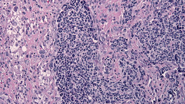
 Histology Glioblastoma with primitive neuroectodermal tumor (PNET) component, HE stain, x100 magnification. Source: Wikimedia Commons.
Jensflorian, via Wikimedia Commons
Biopsy, or physical removal of tissue, followed by a pathologists evaluation of a prepared slice of the tissue is a standard of care in the diagnosis of many diseases. But emerging methods that take an "optical biopsy" offer a new approach -- basically taking the microscope to the tissue rather than the tissue to the microscope. Dr. Arthur Gmitro, Department Head of Biomedical Engineering, describes research at the University of Arizona on the development and use of a confocal microendoscope that takes detailed in-situ images of tissues in real time to allow faster and potentially earlier diagnosis of cancer at the cellular and even molecular levels.
IN THIS EPISODE
Arthur Gmitro, Ph.D, Professor and Head of Biomedical Engineering, Professor of Medical Imaging and Optical Science and Co-Director of the Cancer Imaging Center at the U of A's Cancer CenterLeslie Tolbert, Ph.D, Regents' Professor in the University of Arizona's Department of Neuroscience
MORE: Arizona Science

By submitting your comments, you hereby give AZPM the right to post your comments and potentially use them in any other form of media operated by this institution.